Sunday, March 11, 2007

It's tough creating a model of neonatal skin!!


Making a Neonatal Skin model:

· Firstly we have an approximate skin model from “Skin Optics Summary “ at the website of Oregon medical laser institute.
· This gives us the approximate thickness of each skin layer as follows but it is merely for the sake of a descriptive convention (it says so in the article)[1]
· Thus we refer another article to find out a more in detailed skin optics model [2]
· In this article the thickness of skin layers is widely divergent from those assumed in the Skin Optics Summary (the thickness of the epidermis is 60 um in Skin Optics Summary but 0.027-0.15mm in the Study by the group in Univ.Waterloo)[2][1]
· The skin of a premature neonate is 40-60% less than the skin of an adult – [4]
· The Average thickness of the epidermis is 3 mm and that of the dermis is between 50 and 150 um. [3]
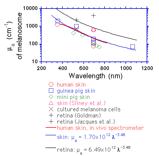
· From the above paper, it was also found that in dark skinned subjects, a higher absorption of melanin causes a reduction in reflectance values, although the overall reflectance pattern remained the same.[3]
· The average error was 0.12%, observed from the arm of a brown skinned subject. [3]
· Moreover, here we are considering the forehead of the neonate, so we first consider the thickness of the dermis and the epidermis in the forehead of an adult
· This is given by an epidermal thickness of mean value 0.253 and standard deviation 0.056[5]
· The dermal thickness is given by a mean value of 1.803 and a standard deviation of 0.252 [5]
· The thickness of adult skin is reported in the literature to vary according to subject and site, with the mean values reported between 0.9 and 1.3 nm [6].
· The twenty neonatal post mortem skin samples obtained for in vitro optical property measurements had a mean thickness of 888 .mu.m, and a standard deviation of 301 .mu.m (Section 2). The thickness of these skin samples was found to have only a weak correlation with gestational age of the neonate, as seen in FIG. 31. This relationship is: Skin thickness =22.5+25 (maturity)[6]
· Note: In the above equation, skin thickness is given in meters, and gestational maturity in weeks.
· In general the thickness of skin is modeled as being infinite
· This is justifiable as long as the wavlength of light is much greater than the thickness of the
· Finally the two models seemed too divergent to be integrated into a single one, thus two separate models were developed: the first being a multilayer model using the adult skin data and the second being a single layer simulation that approximates the epidermal and dermal scattering measurements
· Index of References
· 1-“Skin Optics Summary “, Oregon medical laser Center News, 1998,by Steven L.Jacques
· 2- “A study on Skin Optics” by Aravind Krishnaswamy and Gladimir V.G.Baranoski from the Natural Phenomena Simulation Group, School of Computer Science, University of Waterloo, Canada
· 3 - Electronic biopsy for skin cancer detection, by G.Florence Sudha and T.Ganesa Palnivelu of Pondicherry Engineering College in Current Science, Vol 87,No.5, 10 September 2004
· 4- Course on Neonatal Skin Care by the University of Chicago Children’s Hospital
· 5-Seventeen-point dermal ultrasound scoring system—a reliable measure of skin thickness in patients with systemic sclerosis ,T. L. Moore1, M. Lunt3, B. McManus2, M. E. Anderson1 and A. L. Herrick1,3
· 1University of Manchester Rheumatic Diseases Centre and 2Radiology Directorate, Hope Hospital, Salford M6 8HD and 3ARC Epidemiology Unit, University of Manchester M13 9PT, UK. ,Oxford Journal, Rheumatology 2003; 42: 1559-1563
6-www.freepatentsonline.com –Patent no. 5353790:Method and Apparatus for optical measurement of bilirubin in tissue

Friday, March 9, 2007

Patents,Monte Carlo and Phantoms

I have stumbled onto another remarkable resource- www.freepatentsonline.com/ -
There is a really cool patent for the detection of neonatal jaundice in there,particularly since it provides me with practical information on the construction of the tool.
It also has some formulae that address the minor variations in neonatal skin that occur due to gestational maturity(age after LMP of mother) and other factors such as varying skin thickness etc.
Right now,though,we're still developing the hardware and writing the Monte Carlo Models.
There is some good news,though.After meeting with a professor from the Indian Institute of Technology(Dr.Manivannan) and emailing some other experts in the area
we came to the conclusion that preparing optical phantoms may not be a very good idea at this stage.
Primarily this is because it would just take too long to import the components(Intralipid and other substitutes) at this stage.
So we are directly moving into testing the device on neonates since this seems more important than making the phantoms.The good news is that we have contacted a neonatal specialist who has said that we could test our device on 80-200 children in the ward at Isabel's hospital,over a period of two months.
We will get back to the phantoms once our testing is complete and we have a proper working instrument,by which time it could be used for fine tuning and calibration.
We have been continuously amazed by the people who we have asked for help-they have helped us in the form of their thorough research and practical advice,laboratory equipments that they have offered to let us use,and just words of encouragement.
With an attitude of gratitude,
=)

Monday, February 19, 2007

Classes on Biomedical Optics:Class 1-Radiative Sources


The Oregon Graduate Institute (OGI) has been an amazing source of information throughout the simulation part of this project.
Here is the link to their course on Biomedical Optics:
http://omlc.ogi.edu/classroom/ece532/
From what I have understood, this course deals with how light is transported through a biological tissue.I have summarised it to make it clear to myself:
Class 1:Radiative Sources
If we put a power source in a box,and capture all the energy radiating from it,the rate of increase of the box's energy,per second, is its radiant power.
If we leave it on for a certain time,the total power output gives its radiant energy.
They have also given the example of Laser immersed in black water.
Note to myself:Maybe I could do the same kind of experiment to measure the radiation from the LED source we are going to use.

On the other hand,Radiant Intensity is the power of a source radiating energy into a cone in a particular direction, and it gives the power radiated per unit solid angle.
For instance,if we beam a flashlight onto a wall,the radiant intensity is the
power radiated x (area it falls on)/(total area).
In this case the total area is 4*pi*R^2 and the area it falls on is A.

Similarly,the radiant power per unit surface area is known as the Irradiance.

The Fluence rate,which is similar,refers to the radiant power that is present per unit cross-sectional area of a sphere.

The opposite of irradiance is given by radiant exittance which is the amount of light leaving a surface area.

A more difficult concept is Radiance, which gives the amount of light per unit solid angle(very small),per unit area(again limiting case).

Finally,they have discussed how light is collected.
Firstly,normal laser light can be collected via an aperture,and the irradiance is measured as an exponentially decreasing function of the central axis of the beam.
Secondly,an aperture is used to collect light from conical surfaces.
Thirdly,an integrating sphere is used to collect light.
Finally,the manner in which light is collected and guided by an optical fiber is explained.
An isotropic collector,which collects light with equal efficiency from all directions is also explained.
That's all with the first lecture.
When I study the next lecture , I'll be plunging deeper into biomedical optics.

Absorption Coefficient of Skin Melanosomes

The very first step to simulating the skin is to find out the value of absorption coefficent for the different layers of the skin.
This link provides an idea of the calculation of the absorption coefficient of melanosomes in the skin.
http://omlc.ogi.edu/spectra/melanin/mua.html
Further,they have provided a diagram that illustrates the variation of absorption coefficient of melanosomes for different types of skin,i.e. for human skin,guinea pig skin and the retina.
http://omlc.ogi.edu/spectra/melanin/jacques.mcauliffe.gif
There were 2 very broad conclusions I could make from this
1.The average value of absorption coefficient of melanosomes in human skin is
1.7 x 10^12 x [(lambda*)^-3.48]
*lambda is the wavelength of light used
2.The absorption coefficient of Melanosomes decreases with increasing wavelength of light,thus for lower wavelengths,eg.blue,the absorption will be more.
Two points need to be taken very important note of
1.We are only discussing Melanosomes,and not the total skin characteristics in the above case.
2.The units used are cm-1

Saturday, February 17, 2007

The discovery of SlideShare & the first review presentation

Thanks to Nipun Mehta the tech-wizard,(incidentally the only person in Silicon Valley to be compared to Gandhi (!)) and his fabulous organisation Charity Focus, I found out accidentally about a free,simple,easy to share software called Slideshare.
Nipun has a dream that the Internet will be converted into an Innernet,and it will be a tool where ideas,and not money, enjoy the place of being the primary currency.
The discovery of Slideshare and the no-fuss user interface offered has made me decide that all my presentations from now on will be uploaded using this software.
It never ceases to amaze me how these topnotch companies are able to offer their software free,but I, for one,am not complaining!
As a student, every day I find a new reason to confirm my hunch- that it is really not an exaggeration to say that the Internet opens up our horizons.
In addition,I think Slideshare was started by a bunch of Indian entrepreneurs and they have a team based in New Delhi.
Happy Slide-sharing ! :)

First Review posted

The first project review presentation can be seen here:
http://ideaz.myftp.org/nnj.ppt
Our thanks to Aditya Rajan for his generous domain hosting!

Tuesday, January 16, 2007

Project FAQ's

  • Who comprises the project team?
  • Ajaay Ravi,student of 4th Year Engineering,ECE Department,SRM Institute of Science and Technology
  • Avantika Vardhan ,student of 4th Year Engineering,ECE Department,SRM Institute of Science and Technology
  • Most importantly,our project guide is Mrs.Shanthi Prince,Assistant Professor in the ECE Department of SRM Institute of Technology
  • Mrs.Shanthi Prince is a postgraduate from the Indian Institute of Technology,Madras and is currently pursuing her Ph.D from SRMIST
  • Why is this project being executed?
  • This project is the final-year undergraduate thesis as per the requirements of SRM Institute of Science and Technology for the completion of the B.Tech course in Electronics and Communications (ECE)
  • What is the project title?
  • Currently, our project has been titled "Screening Tool To Detect Neonatal Jaundice by Means of Using Optical Sensors."
  • What are the objectives of this project?
  • The main objective of this project is to create a tool to detect Neonatal Jaundice by means of optical sensor based instrumentation.
  • We also have some secondary objectives, such as:
  • Creating a Monte-Carlo simulation using Matlab that enables us to enhance our accuracy and verify our results
  • Performing Technology-Transfer to make our instrumentation viable for practical use
  • Collecting data samples that test for the validity of the screening tools amongst a cross-section of skin characteristics such as colour.
  • Why did you choose this specific project?
  • India is a developing country with a strikingly high rate of infant mortality. Although jaundice is not a primary reason for this problem, children with jaundice often develop fatal complications.In addition, the rural areas and even urban hospitals are ill-equipped in terms of jaundice diagnosis.For instance, in the whole of Chennai, only Apollo Hospitals has a neonatal jaundice diagnostic lab.
  • Jaundice at the time of birth could result in brain diseases such as kernicterus later on.Our country does not have the health care facilities to help the common man deal well with this dangerous condition
  • Jaundice diagnosis by screening is an exciting area in biomedical engineering and we wish to tailor the existing solutions to suit Indian skin-colors and conditions
  • Invasive procedures expose the child to the risk of trauma,infection and deadly killers such as AIDS if the needles are not properly disinfected and used.Thus, we chose to develop a non-invasive procedure
  • What areas does your project deal with?
  • Our project is interdisciplinary in nature,i.e. it deals with and requires a knowledge of varied branches of study,as stated below.
  • Electronics and Instrumentation : This will deal with the basic part of constructing the instrumentation for the jaundice screening tool
  • Electro-Optics: Since the basis of the sensors used is optical, the branch of electro-optics is central to the project
  • Biomedical Engineering: This field is required for applying the knowledge of the two fields mentioned before, for the specific application in the biomedical field
  • Skin Histology,Anatomy and Dermatology: It is important to know the properties and structure of the skin to create skin samples and design a simulation of skin structure
  • Jaundice - related Medical Science: We have tried to understand the mechanisms involved in bilirubin production which leads to jaundice in order to design the instrumentation correctly.
  • Neonatology: The screening tool we have devised is uniquely designed for neonates.i.e.newborn babies.Thus the data related to skin absorption spectra and skin characteristics, and the intensity of light that the baby will withstand,all make it different from screening jaundice in general.
  • What is the timeline associated with your project?
  • We have already had a Zeroth Review ( the Review proceedings will be posted here) in the end of November
  • Our First Review is to be held on 24th January
  • Our Second and Third Reviews will be held in the end of the months of February and March respectively
  • We may also have a Final Examination in which External Evaluators will award marks to our project
  • Could you give us a brief overview of the instrumentation associated with your project?
  • We are basically using an LED(Light emitting Diode) as a light source which illuminates the skin of the newborn via optical fiber probes
  • The output i.e. absorption and reflectance data are measured by means of instrumentation consisting mainly of a photodiode and an operational amplifier circuit
  • The output will be measured using a reflectance meter built by us using prevalent laboratory based calibration techniques
  • Does your project involve collecting experimental data?
  • Yes,in our final phase we plan to test our instrument on new born babies who are normal and also those with varying degrees of jaundice
  • This will be done with the help of hospitals and testing laboratories around Chennai
  • Will the experimentation be risk free?
  • Fortunately,the main advantage of this project is that the instrumentation is completely risk free
  • For instance, the high power laser-based instrumentation that is used for adults is replaced in our project by an LED light source with very low levels of light intensity-similar to a flashlight or torch